18
Jan
Surgical Microscope in Neurosurgery: Not Luxury but Mandatory!
Introduction
Originally a radical concept, the surgical microscope has become an indispensible tool since the first recorded use of a lighted binocular microscope during an operation in 1922. The simple purpose of this microscope is to improve the surgeon’s view. The basic components of microscopy are magnification, resolution and illumination.
History
The rise of this key surgical tool reflects advances in understanding the principles of optics and vision that have occurred over centuries. The development of reading spectacles in the late 13th century led to the construction of early compound microscopes in the 16th and 17th centuries by Lippershey, Janssen, Galileo, Hooke and others. Leeuwenhoek’s simple microscopes of this era offered improved performance over his contemporaries’ designs. By the late 19th century, Carl Zeiss and Ernst Abbe ushered the compound microscope into the beginnings of the modern era of commercial design and production. The introduction of the microscope into the operating room by Nylen in 1921 initiated a revolution in surgical practice that gained momentum throughout the 1950s with multiple refinements. Kurze is credited with the first application of the microscope to neurosurgery in 1957.Gazi Yasargil made several revolutionary improvements in the design of the operating microscope and is regarded as the “Father of Microneurosurgery”.
Development of neurosurgery
Three technological advances changed the face of neurosurgery: cerebral localization theory, antiseptic/aseptic techniques and anaesthesia. Modern Neurosurgery era is Post- Harvey Cushing. The three technological advances cited have continued to evolve, but neurosurgical practice has also changed enormously and more rapidly in relatively recent times as a result of the operating microscope and the phenomenal advances that have occurred in imaging.
Why Microscope
It has always been a major object of surgery to subject patient to the minimum of strain. One important factor which enables surgeon to operate with a sure, unhesitating hand is a clear recognition of the details of the anatomical structure on which he is working. The brain and the spine are among the most complex parts of the human body and pose a challenge both to the surgeon and to the techniques applied. Operating microscopes help to make structures easier to differentiate, so that operations can be done with maximum precision, leaving the surrounding tissues with little or no harm.
Spine Surgery
By completing the operation through small incisions, the surgeon can safely work on the spine without disturbing normal tissue. As a result, patients are less likely to develop complications, recover sooner and can quickly return to normal activities. Traditional surgical methods for delicate operations require incisions longer in length.
In recent years, technological advances have allowed more back and neck conditions to be treated with a minimally invasive surgical technique.
Benefits of minimally invasive surgical technique in Spine include
- Smaller incisions
- Reduced blood loss
- Less pain
- Less soft tissue damage
- Reduced muscle retraction
- Decreased postoperative narcotics
- Shorter hospital stay
- Faster recovery
- Quicker return to work and activities
Brain Surgery
Now routinely used in almost all intradural operative procedures in the brain. Its use has resulted in smaller wounds, less postoperative neural and vascular damage, better hemostasis, more accurate nerve and vessel repairs and surgical treatment of some previously inoperable lesions.
Benefits of minimally invasive surgical technique in Brain include :
- Permitting neural and vascular structures to be delineated with greater visual accuracy
- Deep areas to be reached with less brain retraction and smaller cortical incisions
- Bleeding points to be coagulated with less damage to adjacent neural structures,
- Nerves distorted by tumor to be preserved
- Enabling anastomosis and suturing of small vessels and nerves not previously possible to be performed
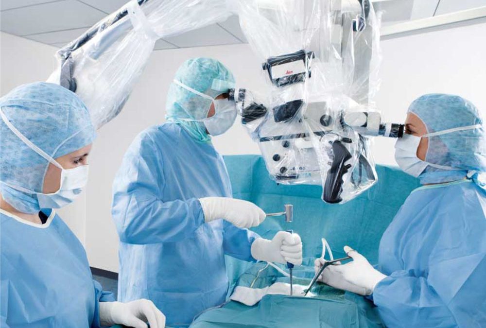
Components of a microscope are
The eyepiece
This consists of a cylinder containing two or more lenses to bring the image into focus for the eye and is located at the observer end of the microscope tube.
Objective lens
This is the cylinder containing one or more lenses to collect light from the operative field and is located at the end closest to the operation site.
The illumination source
This is usually connected to a 100 to 150 watt halogen or Xenon light source via a fiber optic cable and is adjustable. In most operating microscopes, this is achieved by coaxial illumination which means that the light is transmitted through optical system rather than shone onto the operating site externally. Coaxial illumination is superior since it reduces shadows and provides more homogenous and intense illumination of the object.
Head
This is the part with the optics and illumination. The magnification can be changed either manually by adjusting a knob on the head or in some models via a foot pedal. The head can be inclined at an angle, straight or adjustable.
Arm
Heads are attached to an arm of variable length which allows it to be maneuvered into the correct position.
Base or mounting
Microscopes are mounted on a sturdy base which makes it portable. However other options include floor or wall mounting and even a table clamp which allows it to be mounted on a table top.
Advances in Micro-Neurosurgery
The foot controls of the surgical microscope not only adjust the focus and magnification, they are often able to control the movement of the surgeon’s head. When assistants are involved in the surgery they may have a second viewing lens to observe the procedure. Sometimes as many as three assistants are able to view through the same scope. This allows the assistants to be involved in the surgery on a real-time basis. Very often the surgical microscope will have a video display attached and other medical personnel in the operating room are able to keep an eye on the surgery as well.
With the development of these sophisticated devices it is now possible to reattach limbs, perform surgery on the spine and brain and reconnect minute blood vessels. All the surgical microscopes have superior illumination mechanisms, as well as good visual depth of field. This allows the surgeon to continue operating without having to constantly readjust the focus. The microscopes can come with back up light systems, in case of failure in the primary bulb during a critical phase of the surgery.
- Microscope head has a camera to document the procedure on an SD card or DVD as well as to allow colleagues to view the operation in real time.
- Improved handling and practical operation of the surgical microscope.
- 2 or more Surgeons can work together.
- Fluorescence Microscopy
Cost-efficiency of Microscope
The Initial investment on a Surgical Microscope can be exorbitant. But the cost is offset by the benefits to the patients and satisfaction to the operating surgeon. Since this type of microscope is quite expensive, purchasing one that can be used by a variety of medical specialties is a cost effective solution.
Conclusion
The operating microscope is a fixture of modern surgical facilities and it is a critically important factor in the success of many of the most complex and difficult surgical interventions used in medicine today.
Today, Neurosurgery cannot be conceived without operating microscopes.
Dr. Viswanathan Iyer is a Consultant Brain and Spine surgeon. His special interest is Neurovascular Diseases, Stroke and Neuro-intervention.
He is a Consultant at Fortis Hospital, Bethany Hospital, Kohinoor Hospital etc.